- Total knee arthroplasty is associated with severe post-operative pain in up to 36%
- Severe pain is associated with:
- Reduced patient satisfaction
- Delayed mobilisation and prolonged hospital stay
- Increased healthcare costs
- Increased rates of chronic pain
- Early mobilisation is central to ERAS e.g. physiotherapy reduces length of stay by ~1.8 days
- Femoral (± sciatic) nerve blocks provide excellent analgesia but cause quadriceps weakness (and foot drop), delaying mobilisation and increasing falls risk
- The goal is analgesia that preserves quadriceps strength, while avoiding the side-effects of systemic analgesics
- Achieved by targeting the sensory (articular) terminal branches around the knee rather than the motor trunks proximally
- Delivered as part of multimodal analgesia
- Can facilitate same-day discharge in selected patients
- Low concentrations of LA are preferred, e.g. 0.2% ropivacaine, 0.125-0.25% (levo)bupivacaine
Motor-Sparing Blocks for Knee Surgery
Motor-Sparing Blocks for Knee Surgery
This topic is probably outside the purview of the FRCA exams.
Resources
- Motor-sparing regional anaesthesia for total knee arthroplasty: a narrative and systematic literature review (British Journal of Anaesthesia, 2025)
- Anaesthesia and analgesia for total knee arthroplasty (BJA Education, 2026)
- Adductor canal blocks for postoperative pain treatment in adults undergoing knee surgery (Cochrane, 2019)
- The nerves of the adductor canal and the innervation of the knee: an anatomic study (Regional Anesthesia & Pain Medicine, 2016)
Femoral triangle and adductor canal
- Femoral triangle borders:
- Superior: inguinal ligament
- Medial: adductor longus
- Lateral: sartorius
- Apex: where the medial borders of sartorius and adductor longus intersect; identifiable on ultrasound
- Adductor canal: runs from the apex of the femoral triangle to the adductor hiatus
- Roof: vastoadductor membrane and sartorius
- Lateral: vastus medialis
- Medial: adductor longus and magnus
- Adductor hiatus: gap in adductor magnus through which the femoral vessels pass posteriorly to become the popliteal vessels
- Contents: femoral artery and vein, saphenous nerve, nerve to vastus medialis (NVM) ± posterior and anterior branches of the obturator nerve and medial femoral cutaneous nerve
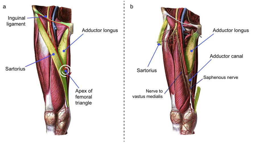
(a) Borders and apex of the femoral triangle; (b) sartorius reflected to show the adductor canal, saphenous nerve and NVM. Adapted from Motor-sparing regional anaesthesia for total knee arthroplasty: a narrative and systematic literature review
Knee joint innervation
- Complex, variable and inconsistently described in the anatomical literature
- Follows Hilton's law: a joint is innervated by articular branches of the nerves supplying the muscles that move it
- Derived from the femoral, obturator and sciatic nerves (lumbar and sacral plexuses) and is usually simplified into anterior and posterior compartments, then quadrants
Anterior knee
- Femoral nerve: divides below the inguinal ligament into:
- Anterior division: motor to sartorius + anterior femoral cutaneous nerves (AFCNs i.e. intermediate and medial femoral cutaneous nerves), supplying the skin of the anterior knee
- Posterior division: motor branches to quadriceps, which divide in the iliopectineal fossa (a proximal injection here causes weakness) and their terminal branches become sensory to the knee
- Femoral sensory contributions:
- NVM: superomedial quadrant as usually gives rise to the superomedial genicular nerve
- Nerve to vastus intermedius: medial and lateral articular branches to the suprapatellar bursa and both superior quadrants
- Nerve to vastus lateralis: superolateral quadrant
- Infrapatellar branch of the saphenous nerve (arises after the saphenous nerve exits the adductor canal): inferomedial quadrant and skin
- Sciatic contributions (despite their posterior origin, these mostly supply anterior structures):
- Superolateral genicular nerve: sciatic or common peroneal nerve
- Inferolateral genicular nerve: common peroneal nerve
- Inferomedial genicular nerve: tibial nerve or popliteal plexus
- Recurrent peroneal nerve: common peroneal nerve
- Genicular nerves supply the periosteum and anterior capsule, they travel with the genicular arteries, which act as ultrasound landmarks
- Obturator contribution is variable and inconsistent
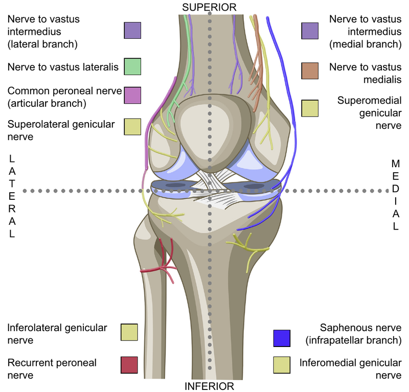
Simplified innervation of the anterior knee. Adapted from Motor-sparing regional anaesthesia for total knee arthroplasty: a narrative and systematic literature review
Posterior knee
- Supplied largely by the popliteal plexus, formed from articular branches of the:
- Tibial nerve (the consistent contributor)
- Posterior branch of the obturator nerve; runs in or below the adductor canal and passes through the adductor hiatus
- Common peroneal nerve
- Innervates the posterior capsule and intra-articular structures
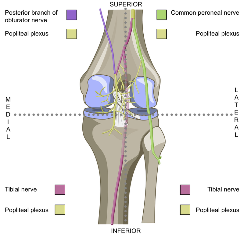
Simplified innervation of the posterior knee. Adapted from Motor-sparing regional anaesthesia for total knee arthroplasty: a narrative and systematic literature review
Matching blocks to territories
| Territory | Block |
| Anteromedial capsule (saphenous nerve, NVM) | Adductor canal/femoral triangle block |
| Anteromedial skin and incision | Anterior femoral cutaneous nerve block |
| Anterior capsule and periosteum | Genicular nerve blocks Nerves to vastus intermedius and lateralis |
| Posterior capsule | iPACK (or surgical LIA) |
- Surgical infiltration of large volumes of dilute LA into the periarticular tissues
- At least two stages: posterior capsule before component insertion, then anterior, medial, lateral and subcutaneous tissues afterwards
- Typical dose ~400mg ropivacaine for unilateral TKA (up to 800mg bilateral)
- Additives include adrenaline, NSAIDs (e.g. ketorolac), steroids, opioids, α2-agonists, antibiotics and TXA
- Mechanism: direct sensory block, dilution of inflammatory mediators, direct anti-inflammatory effect and a depot providing ongoing systemic analgesia
- Recommended by ERAS, PROSPECT and NICE and is considered the benchmark against which motor-sparing blocks are compared
- LIA and peripheral nerve blocks are not mutually exclusive as adding blocks can reduce the total LA dose in at-risk patients
| Advantages | Disadvantages |
| Simple, quick and accessible | Heterogeneous technique, volumes and additives |
| Opioid-sparing; motor-sparing | Weak evidence base for optimal mixture and site |
| Shorter stay, better range of movement at 24hrs and higher satisfaction vs. placebo | High LA doses, often above manufacturer limits; risk of LAST |
| No additional anaesthetic time | Off-licence use of additives |
| Useful when nerve blocks are impractical | Single-shot; no role as a rescue technique |
LAST risk
- Measured free ropivacaine concentrations usually remain below toxic thresholds, but some patients approach or exceed them
- Time to peak plasma concentration is unpredictable: as early as 20mins with plain ropivacaine, delayed to 2-6hrs by adrenaline
- Peak levels may therefore occur after discharge from recovery which is relevant when planning monitoring and rescue blocks
- Reduce doses at extremes of age and weight, in females and with organ dysfunction
Subsartorial blocks: femoral triangle and adductor canal
- Historically confused with many trials describing 'adductor canal blocks' which were in fact mid-thigh femoral triangle blocks
| Femoral triangle block | Adductor canal block | |
| Injection site | Proximal to the apex of the femoral triangle | Distal to the apex, proximal to the adductor hiatus |
| Nerves blocked | Saphenous nerve, NVM ± medial femoral cutaneous nerve | Saphenous nerve ± posterior branch of obturator nerve |
| Volume | ~15ml | 20-30ml (mid-thigh) |
| Quadriceps weakness | Higher risk (closer to motor branches) | Lower risk |
- Technique:
- Supine, 'frog-leg' position with high-frequency linear probe transverse on the medial thigh
- Identify the femoral artery deep to sartorius and slide distally to find the apex of the femoral triangle
- In-plane, lateral to medial, beneath sartorius towards the artery; 5-20ml LA adjacent to the artery depending on desired effect
- Nerve stimulation can identify the NVM, either to block it selectively or to avoid injuring it
- Evidence:
- Femoral triangle, proximal and distal adductor canal blocks produce similar analgesia and mobility
- Comparable analgesia to femoral nerve block with better preservation of quadriceps strength
- Comparable analgesia to LIA and adding a block to LIA gives superior analgesia to either alone
- Covers only the anteromedial knee, hence the need for complementary techniques
- Catheters: conflicting meta-analyses with no clear benefit in the first 24hrs but may improve pain at 24-48hrs
- Not advised routinely, but consider in opioid-tolerant patients and as part of rescue analgesia
- The NVM contributes more to anteromedial capsule innervation than the saphenous nerve, so blocking both is desirable
- The muscular branch of the NVM enters vastus medialis just below mid-thigh, and blocking it may explain occasional quadriceps weakness after adductor canal block (BJA, 2021)
Anterior femoral cutaneous nerve block
- The AFCNs supply the anteromedial thigh and knee skin and are usually missed by femoral triangle and adductor canal blocks
- The medial femoral cutaneous nerve may be caught by a proximal femoral triangle injection
- The intermediate femoral cutaneous nerve branches more proximally and needs a separate injection
- Technique: linear probe transverse at the proximal mid-thigh injecting 3-5ml LA into the fat-filled plane superficial to the fascia lata over sartorius
- Adding to a femoral triangle block improves dynamic pain for ~12hrs, incision pain at 24hrs and morphine consumption over 48hrs
Genicular nerve blocks
- Targets: superomedial, superolateral and inferomedial genicular nerves
- The inferolateral genicular and recurrent peroneal nerves are usually avoided due to the risk of common peroneal nerve block and foot drop
- Technique:
- Linear probe longitudinal over the metaphyseal-diaphyseal junction of the distal femur or proximal tibia
- Identify the genicular artery and deposit 4-5ml LA per target onto the periosteum
- Evidence:
- Improve analgesia vs. placebo
- Sdding them to adductor canal block + iPACK more than halved opioid use in the first 24hrs
- Equivalent to high-volume LIA using only 20-40ml LA rather than ~150ml
- Lower doses allow more concentrated LA, additional blocks within safe limits, or bilateral TKA
Nerves to vastus intermedius and lateralis
- Nerve to vastus intermedius: its articular branches run between the femur and vastus intermedius in the distal third of the thigh; LA onto the anterior femoral periosteum just above the patella
- Nerve to vastus lateralis: targeted at mid-thigh, adjacent to the descending branch of the lateral circumflex femoral artery
- 3-5ml LA each may add benefit, but neither has been assessed in isolation in comparative trials
iPACK block
- Infiltration between the popliteal artery and capsule of the knee
- Targets popliteal plexus branches while sparing the tibial and common peroneal nerves
- Cadaveric studies suggest some spread to anterior articular branches too
- Technique:
- 'Frog-leg' position with curvilinear probe transverse over the distal medial thigh
- Trace the femoral vessels distally until the popliteal artery lies posterior to the femur, just proximal to the condyles
- In-plane, medial to lateral, parallel to the femoral shaft until the tip is ~2cm lateral to the artery
- ~20ml (10-30ml) LA injected incrementally as the needle is withdrawn
- Avoid a lateral-to-medial approach due to the risk of common peroneal nerve injury
- Evidence:
- Without LIA: improves analgesia and function for the first 24hrs
- Equivalent or better analgesia than LIA using half the LA dose
- No additional benefit when added to LIA, as both treat the posterior capsule
Selective tibial nerve block
- Avoids foot drop from common peroneal block, but not fully motor-sparing
- Not superior to LIA, and slower mobilisation than iPACK, probably due to variable spread to the sciatic bifurcation
- Superior analgesia to femoral triangle and adductor canal blocks, but quadriceps weakness has removed it from routine ERAS pathways
- The analgesic gap is probably due to missed AFCNs and terminal branches of the nerves to vastus lateralis and intermedius
- Better analgesia and less nausea than opioid PCA, and similar analgesia to epidural and LIA (Cochrane, 2014)
- Remaining roles:
- Rescue analgesia in recovery using a short-acting LA (e.g. 2% chloroprocaine), which improved pain and opioid use
- Lidocaine femoral block for TKA under GA (without spinal) alongside motor-sparing blocks, to reduce recovery pain and opioid use
- Dense femoral ± sciatic blocks for complex revisions, planned delayed mobilisation or severe pre-operative pain
- The optimal combination remains unclear, with evidence strongest for subsartorial blocks and limited for other targets
- PROSPECT: single-shot adductor canal block + LIA is the standard approach
- Anatomically complete coverage would combine:
- Adductor canal/femoral triangle block (anteromedial capsule)
- AFCN block (skin)
- Genicular nerve blocks (anterior capsule)
- iPACK (posterior capsule)
- No RCT has compared all four against LIA although a small retrospective study of a multi-block regimen reduced 48hr morphine use by 68% vs. LIA + adductor canal block
- Reasonable routine options:
- Adductor canal block + LIA
- Adductor canal block + genicular nerve blocks, which may be preferred in those at risk of LAST
- Calculate the maximum LA dose meticulously when combining blocks with each other or with LIA
Special populations
- Most trials exclude extremes of age and BMI, ASA 3-5, organ dysfunction, high-dose opioid use and substance use
- These patients are hardest to manage and stand to gain most from multi-block techniques and catheters
Motor block
- Quadriceps weakness and falls:
- Proximal spread of large volumes to the femoral nerve motor branches
- Direct block of the NVM muscular branch
- Rarely, myositis from spread outside the vastoadductor membrane; profound weakness that resolves over months
- Foot drop from common peroneal nerve involvement:
- Distal spread of adductor canal injectate into the popliteal fossa
- Inferolateral genicular or recurrent peroneal nerve blocks
- Lateral-to-medial iPACK approach or spread to the sciatic bifurcation
Other
- LAST, particularly with combined blocks and LIA
- Vascular puncture or intravascular injection: femoral, popliteal and genicular vessels
- Nerve injury
- Catheter problems: migration, leakage, obstruction, dislodgement and infection