- Glucose is small (molecular weight 180Da) and not protein-bound, so is freely filtered
- The filtered load is directly proportional to plasma glucose concentration:
- Filtered load = GFR × plasma glucose concentration
- E.g. 180L/day × 5mmol/L = 900mmol/day
- 900mmol × 180g/mol ÷ 1000 = ~160g of glucose filtered each day
- Virtually all of this is reabsorbed, so normal urine is glucose-free
Renal Glucose Handling
Renal Glucose Handling
Resources
- Glucose is freely filtered at the glomerulus and, in health, almost entirely reabsorbed in the proximal convoluted tubule (PCT)
- Reabsorption is a saturable, carrier-mediated process, so glucose appears in the urine once plasma glucose exceeds the renal threshold
- This mechanism is the target of SGLT2 inhibitors
Apical membrane
- Glucose is reabsorbed by secondary active transport, coupled to Na+ moving down its concentration gradient
- The gradient is maintained by the basolateral Na+/K+-ATPase
| SGLT2 | SGLT1 | |
| Location | Early PCT | Late PCT (also small intestine) |
| Na+:glucose stoichiometry | 1:1 | 2:1 |
| Affinity / capacity | Low affinity, high capacity | High affinity, low capacity |
| Proportion of glucose reabsorbed | ~90% | ~10% |
Basolateral membrane
- Glucose leaves the tubular cell down its concentration gradient into the interstitium by facilitated diffusion
- This occurs via GLUT2 (early PCT) and GLUT1 (late PCT)
SGLT2 inhibition
- Blocking SGLT2 causes loss of several grams of glucose (and the associated Na+) in the urine each day
- Only ~50% of filtered glucose is excreted, as SGLT1 in the S3 segment upregulates and compensates
- Reabsorption increases linearly with filtered load until the carriers start to saturate
- Renal threshold: the plasma glucose at which glucose first appears in the urine, ~10-11mmol/L
- Transport maximum (Tmax): the maximal rate of tubular glucose reabsorption, ~1.5-2mmol/min (~300-375mg/min)
- Once Tmax is reached, reabsorption plateaus and any further filtered glucose is excreted
Splay
- Glucose appears in the urine at a plasma concentration below that predicted from Tmax, so the titration curve is rounded rather than sharply angled
- This is because:
- Nephrons are heterogeneous, with differing filtration rates and reabsorptive capacities, so some saturate before others
- Carrier binding is not infinitely avid, so some glucose escapes reabsorption before full saturation
Glucose titration curve
- Plotting filtered load, reabsorption and excretion against plasma glucose gives three lines:
- Filtered load: a straight line through the origin
- Reabsorption: follows the filtered load, then curves (splay) and plateaus at Tmax
- Excretion: zero until the renal threshold, then rises in parallel with filtered load
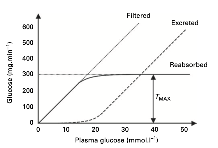
Adapted from Physics, Pharmacology and Physiology for Anaesthetists: Key Concepts for the FRCA
- Glycosuria occurs once the renal threshold is exceeded
- Above Tmax, excretion rises linearly in parallel with filtered load
- Glucose in the tubular fluid holds water osmotically, causing an osmotic diuresis with loss of water and electrolytes
- This explains the polyuria, polydipsia and dehydration of uncontrolled diabetes and DKA
Glycosuria without hyperglycaemia
- Pregnancy: ↑ GFR raises the filtered load, and the renal threshold falls
- SGLT2 inhibitor therapy
- Familial renal glycosuria (SGLT2 gene mutations)
- Proximal tubular dysfunction e.g. Fanconi syndrome, heavy metal toxicity
Altered threshold in diabetes
- In poorly controlled diabetes, SGLT2 expression increases, raising Tmax
- This maladaptive response increases glucose reabsorption and worsens hyperglycaemia
- Conversely, the renal threshold may be raised in the elderly and in diabetic nephropathy, so glycosuria is an unreliable marker of hyperglycaemia